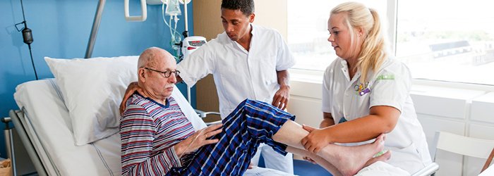
Wound Care in Hospice Settings
The goal of hospice is to promote quality of life with a focus on managing pain. The skin deterioration observed in pressure ulcers or other wounds is a symptom of body systems breaking down.
Even in the final stages of life, good wound care can contribute to physical, psychological and emotional comfort. Wounds can generate feelings of fear, aversion and suspicion of neglect. Poor wound care—or lack of wound care—can be devastating to the patient and family’s experience of death.
Pressure Ulcers in Hospice Patients
Pressure ulcers (bedsores) occur in more than 40 percent of hospice patients. Even with aggressive preventive measures, critically ill patients experience compromised healing response due to impacted muscle cell and immune function, among other factors. For these patients, pressure ulcer formation may be a visual biomarker that the critical illness has overwhelmed the body; skin breakdown is neither preventable nor treatable.
Family members of terminally ill patients may view pressure ulcer formation as a failure on the part of the healthcare staff caring for the patient—or even as their own failing, if they are responsible for providing care. Their emotional response may lead to requests that compromise the patient’s comfort-focused plan of care.
However, in general, prevention and treatment should not compromise the hospice philosophy of providing comfort care. Studies show that when patients are more comfortable in one position due to advanced illness, comfort should supersede preventive measures. If hospice staff deem that routine patient turnings contribute to increased pain, turnings may be suspended.
Other Wounds Common in Hospice
Due to the wide variety of patients and conditions treated in hospice care, you may encounter an equally wide variety of wound types:
- Arterial insufficiency—Often appears as ulcers with black eschar on the lower leg and foot. The skin surrounding the wound appears to be very thin, shiny and usually hairless. The foot may feel cold and appear dusky red or pale.
- Diabetic ulcers—Typically on the plantar surface of the foot and the second metatarsal head. They are usually painless.
- Venous ulcers—Occur in the so-called gaiter area, halfway up the calf and down to just below the ankle. The skin likely feels itchy and appears mottled brown or has black staining and may appear crusty. The legs may become painful with sitting.
- Tumors or fungating lesions—Occur most often in cancer of the breast, but may occur with other types of cancers, including head and neck, malignant melanomas and sarcomas. The lesion may be a small crusted area or a large ulcerated area with profuse exudate and capillary bleeding. They are often disfiguring, distressing and isolating. Odor and exudate management may be a particular problem in this type of wound.
Risk Factor Assessment and Prevention
Risk assessment upon admission is crucial and must include a full body check. Pressure ulcers are caused by intrinsic and extrinsic factors, including immobilization, cognitive deficit, inability to verbalize discomfort or numbness, chronic illness, aging and poor nutrition.
Once risk factors are established, preventing wounds from occurring is the best practice. Prevention measures include, but are not limited to, inspecting the skin and monitoring for proper moisture control. Proper positioning, transfer techniques and nutrition are essential for the comfort of the patient.
Avoid pressure on the heels and bony prominences of the body, and use positioning devices whenever feasible. As always, remember to document the condition of the skin after assessment.
Staging Wounds
The following wound stages are established by the National Pressure Advisory Panel:
- Stage I—The skin is intact with non-blanchable redness of a localized area. Darkly pigmented skin may not have visible blanching, but its color may differ from the surrounding area. The area may be painful, firm, soft, warmer or cooler as compared to adjacent tissue.
- Stage II—Look for partial-thickness loss of dermis presenting as a shiny or dry shallow open ulcer with a pink wound bed, without slough or bruising. It may also present as an intact or ruptured serum-filled blister. Stage II does not describe skin tears, tape burns, perineal dermatitis, maceration or excoriation.
- Stage III—Indicates full-thickness tissue loss. Subcutaneous fat may be visible, but bone, tendon or muscle is not exposed. Slough may be present but does not obscure the depth of the tissue loss. There may be undermining and tunneling. The depth of a stage-III pressure ulcer varies by anatomical location.
- Stage IV—Full-thickness tissue loss with exposed bone, tendon or muscle. Slough or eschar may be present on some parts of the wound bed. This wound often includes undermining and tunneling. The depth of a stage-IV pressure ulcer varies by location. Stage-IV ulcers can extend into muscle and/or supporting structures, making osteomyelitis possible.
- Deep-Tissue Injury—A purple or maroon localized area of discolored intact skin, or a blood-filled blister due to damage of the underlying soft tissue from pressure and/or shear. The area may be preceded by tissue that is painful, firm, mushy, boggy, warmer or cooler as compared to adjacent tissue.
- Unstageable—Full-thickness tissue loss in which the base of the ulcer is covered by slough (yellow, tan, gray, green or brown) and/or eschar (tan, brown or black) in the wound bed. Until enough slough and/or eschar is removed to expose the base of the wound, the true depth, and therefore stage, cannot be determined.
Basic Principles of Wound Care
The first step in determining a successful wound plan of care involves establishing the patient’s prognosis, condition and potential for wound healing. For instance, a less aggressive approach will be necessary for someone in the final days of life, or when it is evident that healing is not realistic.
It’s important to set appropriate goals based on the prognosis, condition and potential for healing. Goals for hospice patients may include:
- Preventing complications of the wound, such as infection or odor
- Preventing additional breakdown of the skin
- Minimizing harmful effects of the wound on the patient’s overall condition
When developing a care plan for a patient, it is important to factor in their status and desires. Interventions should be re-evaluated every two weeks to determine whether the plan is still appropriate. A wound’s failure to heal does not necessarily imply that other measures are more appropriate, or that all possible approaches should be attempted. Instead, focus on developing the best option for the patient based on their present condition and their desires.
A basic wound care plan will incorporate these elements:
- Cleansing debris from the wound
- Possible debridement
- Absorbing excess exudate
- Promoting healing
- Treating infection
- Minimizing discomfort
In hospice care, the pain, appearance, odor and perceived implications of wounds can be incredibly distressful for the patient and family. To stay in alignment with hospice’s mission of comfort care, the entire hospice team must operate with vigilance, assessing and documenting wounds and condition changes in detail, maintaining proper preventive measures and developing plans of care that consider the patient’s needs and desires.
By reducing the occurrence and severity of wounds and mitigating their effects, the hospice team contributes to quality of life and facilitates a peaceful death experience for both patient and family.
See upcoming continuing education opportunities through VITAS.


Check Hospice Guidelines
Get diagnosis-specific guidelines in our hospice eligibility reference guide.
Hospice Guidelines by Diagnosis Refer Your Patient